

Automated Cell Counting for Malaria Detection
Executive Summary
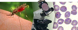 The major tool for diagnosing malaria is microscopy. Hundreds of millions of blood slides are inspected under the microscope every year for parasites causing malaria. This is a tedious and error-prone manual process, whose success depends on the experience of a microscopist.
To reduce the diagnostic burden, the National Library of Medicine (NLM), part of the National Institutes of Health, has developed software that can count parasite-infected and uninfected red blood cells using automatic image analysis and machine learning algorithms developed by computational scientists at NLM and the University of Missouri. With funding from the HHS Secretary's Ventures Fund, this software has been ported to Android smartphones. Running on a camera-equipped smartphone attached to a microscope by an adapter, the software screens the field of view for malaria parasites and reports the level of parasitemia to the microscopist.
The software has been trained with more than 200,000 blood cell images, which the NLM team acquired in Bangladesh and have been annotated by an expert microscopist.
The team is now field testing the software in Bangladesh and Thailand, following which the software will be made widely available to other sites.
A project supported by the: HHS Secretary's Ventures Fund
Team Members
Stefan Jaeger (Project Lead), NIH
Sameer Antani, NIH
Hang Yu, NIH
Rick Fairhurst, NIH
Abhisheka Bansal, NIH
Richard Maude, University of Oxford (UK)
Kamolrat Silamut, Mahidol University (Thailand)
Milestones
April 2015: Project received support from the HHS Secretary's Ventures Fund
July 2015: Field system design finalized (smartphone, microscope, and adapter)
August 2015: Begin image acquisition in the field
September 2015: Roadmap for software porting to smartphone finalized
October 2015: Annotation tool development and start of image annotation
December 2015: End of image acquisition
January 2016: End of image annotation
February 2016: First smartphone application prototype developed
April 2016: Patient database added and user feedback incorporated
May 2016: Support from HHS Secretary's Ventures Fund ends
Project Sponsor
George Thoma, Chief, Communications Engineering Branch, Lister Hill National Center for Biomedical Communications, National Library of Medicine, National Institutes of Health
Additional Information
Contributing Partners:
Kannappan Palaniappan, University of Missouri
Ilker Ersoy, University of Missouri